USP
GratXray’s GI-BCT is the first one-stop solution, for efficient, accurate and painless breast cancer diagnosisGratXray Solution
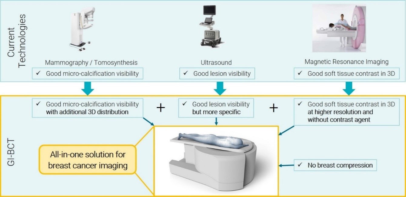
GratXray is developing a disruptive medical device for accurate, reliable and painless breast cancer diagnosis that will cut costs on the pathway of breast cancer care. The uniqueness of our solution lies in the fact that we are the only company integrating grating-interferometry (GI) into a medical device. This allows for the first time to simultaneously acquire absorption, refraction and scatter signals of X-ray at a very high resolution. This cutting-edge technique can be applied to a broad variety of medical applications and has a high potential to revolutionize the whole medical X-ray imaging field. The refraction and scatter signals have so far been considered as noise in conventional X-ray diagnostic and systematically removed from clinical radiographs. However, these new signals allow for significantly improved image quality. The refraction signal refines lesions delineation, while the scattering signal provides micro-structural breast information. Integrated in a breast computed-tomography (BCT) device, this yields in a 3D imaging system providing a comprehensive solution to current breast imaging problems at three different levels: physical and mental health, diagnosis, and economics. Unlike any other competitor, our GI-BCT system not only integrates all the strengths of existing technologies in a single multi-modality device, but thanks to its technology based on grating interferometry, it also provides additional capacities and advantages, as illustrated in Figure 1.
The table shows the impact of GI-BCT on the main stakeholders identified: women (end-users), clinicians (users), breast centers and hospitals (buyers), as well as both, public and private health insurances (society).
- Survival rate
- Anxiety
- Discomfort
- Greater chances of survival by early detection.
- Anxiety and psychological stress are avoided by precise, immediate and reliable results.
- Women are more predisposed going to breast cancer imaging as there is no breast compression.
- Inaccuracy
- Efficency
- Pain
- Fatty as well as dense breast tissue are imaged with same high accuracy.
- GI-BCT allows reliable diagnosis by improving the strengths of conventional technologies and by providing additional diagnostic values such as tumor lesion differentiation, micro-calcification classification, improved soft-tissue discrimination and accurate tissue density assessment.
- It simplifies the results’ interpretation by displaying all the information in a single image instead of uncorrelated data acquired independently.
- A three times faster diagnosis, as compared to FFDM plus US, saves physician time, increasing the patients’ number and therefore multiplying the revenue.
- High costs
- Save time
- Reduce recall rates.
- Accurate results are immediately available saving time of clinicians.
- A single multi-modality GI-BCT system has an acquisition and running cost comparable to that of FFMD and US systems together but it offers additional economic benefits.
- Fewer recalls reduce costs from additional procedures, saving physicians time as well as admin costs.
- As an all-in-one solution, GI-BCT allows to save time, opening the door to new patients and reducing managing costs in patient care and equipment maintenance.
- New clients are attracted by improving the reliability and performance of the imaging system. Breast centers can take care of more patients.
- An early detection of breast cancer leads to reduced treatment costs, thus, cutting costs to the society.